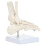
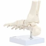
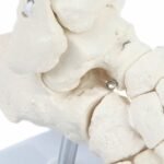
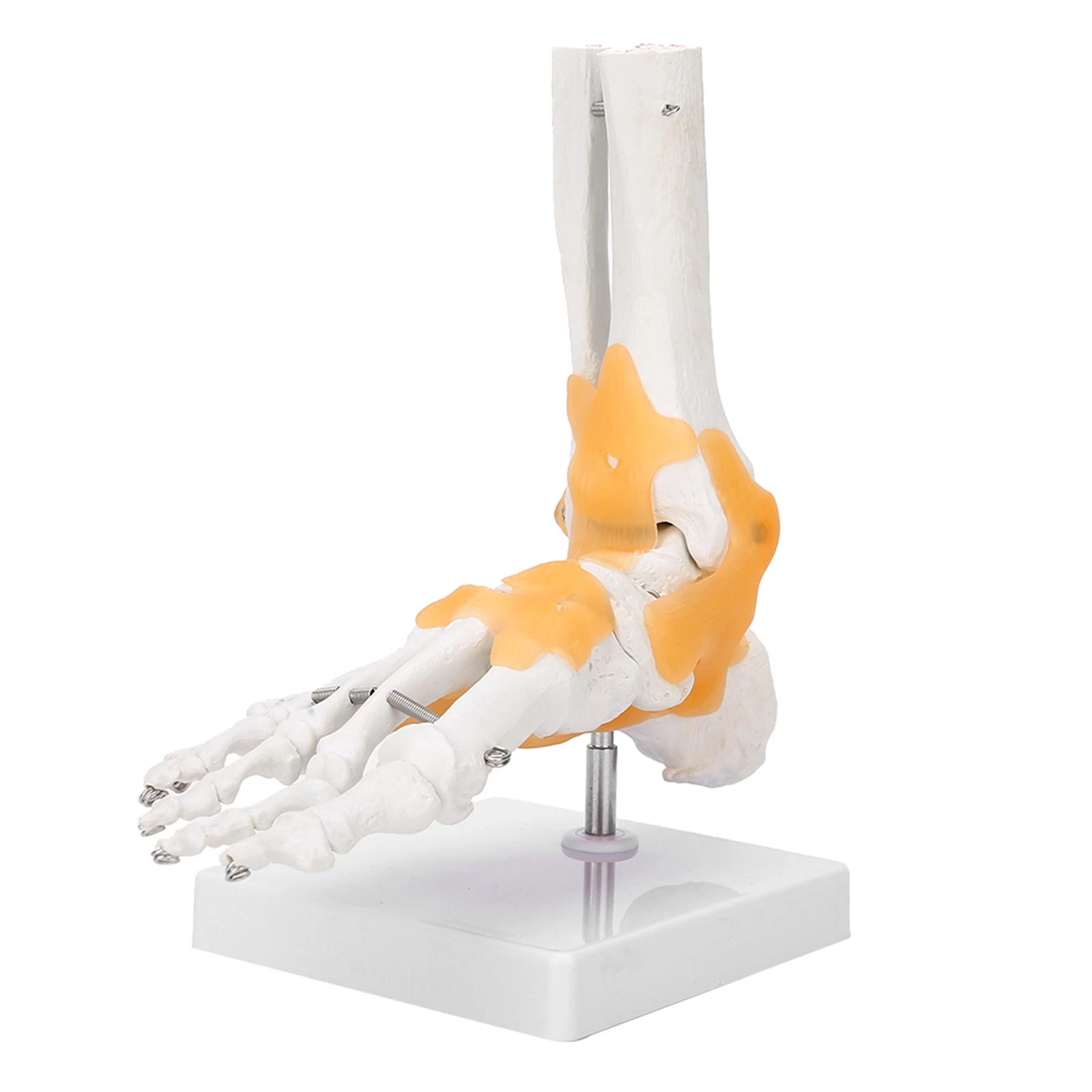
Axis Scientific is pleased to introduce an updated and very detailed model of the human foot. This model offers better access and visibility to all the bones in the foot.
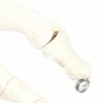
This Axis Scientific anatomy model features a complete human foot with the lower portions of the tibia and fibula bones included. The Bones of the foot are held together with high quality wire which also allow for mobility and flexibility.
This foot anatomy model is great for podiatry students and podiatrists who want to learn more about the bony landmarks and structures of the human foot. The realistic appearance and easy display make this anatomy model great for patient education and understanding of common foot injuries and ailments.
Features:
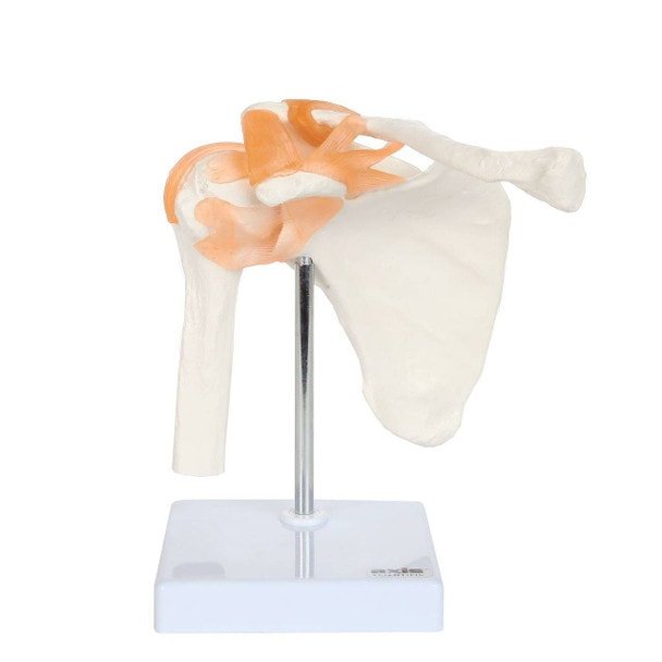
Life-Size Human Knee Joint Anatomy Model
The Axis Scientific Life-Size Knee Joint Model features 18 bones, ligaments, and tendons involved in the knee joint’s anatomy. Cast from real human bones, the model includes the fibula, tibia, patella, femur and the associated bony landmarks and textures.
Our knee joint model offers an anatomically correct representation of the connection between the different bones in the area and how they relate to the anatomy, structure, and stability of the knee. As one of the strongest and most complex joints in the human body, the knee joint can support the body’s weight when upright and help propel the body forward when walking. This anatomy model features the patella (kneecap) which provides articulation to the femur.
Knee Ligaments
This knee joint anatomy model features textured ligaments to provide a better understanding of the connective structures around the bones. The tough elastic tissues are presented prominently on the model to represent a detailed view of the human knee joint’s anatomy.
The human knee joint is susceptible to several injuries. These injuries can be from common, daily events or athletic in nature. The ligaments represented on the model includes the anterior cruciate ligament (ACL) and the posterior cruciate ligament (PCL), both major ligaments that provide stability to the knee joint. Tears and sprain commonly occur in these ligaments, most notably in sports. This knee joint with ligaments model allows for the study of how these ligaments co-exist with the other ligaments, tendons, and bones in the knee.
Sturdy Base Stand
Our knee joint with ligaments model comes with a sturdy base stand, making it perfect for study, display, and transport. The base stand allows the model to be viewed upright for an uninterrupted study of the knee joint anatomy.